Japanese Researchers Link Compromised Synapse-Clearing Ability to Autism
A new study finds that macrophages from individuals with autism have a significantly impaired ability to clear synaptic proteins
Microglial dysfunction has been implicated in autism spectrum disorders (ASD), but limited access to human brain tissue posed challenges in related research. A new study explored how immune cells called monocyte-derived macrophages can serve as models for microglia in ASD. Researchers used these cells to investigate neuroimmune responses, such as synaptic phagocytosis in ASD. These findings could open new avenues for understanding the brain-immune system connection in ASD and identifying potential targets for therapy.
Autism spectrum disorder (ASD) is a complex neurodevelopmental condition in which affected individuals experience difficulties in social communication and exhibit restricted, repetitive patterns of behavior or interests. A growing body of research suggests that neurobiological changes, particularly abnormalities in dendritic spines, tiny protrusions on nerve cells where synapses form, may be a hallmark of ASD. In particular, studies have found an unusually high number of these spines in individuals with autism. This overabundance of synaptic connections could disrupt normal communication pathways in the brain, potentially contributing to the behavioral and cognitive features seen in ASD.
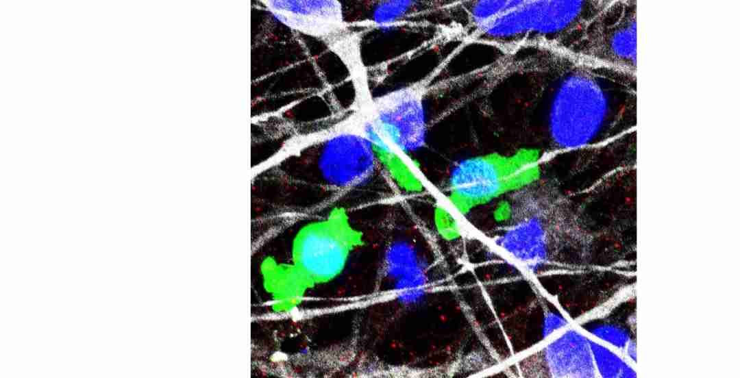
(white: neurites of hiPS cell-derived neurons, green: macrophages from individuals with ASD, red: PSD95, a scaffolding protein in the postsynaptic region, blue: nuclei)
Image License: CC BY-ND 2.0
Under normal circumstances, the brain undergoes synaptic pruning, a process involving the removal of unnecessary or weak synaptic connections to make way for more efficient neural networks. This pruning is crucial during early development and adolescence. Microglia, the resident immune cells in the brain, play a critical role in this process. In addition to defending the brain against infection or injury, they act like gardeners by trimming away excess synapses to help shape healthy neural circuits. However, studying how microglia function in the human brain, especially in individuals with autism, has proven difficult. Unlike in animal models, directly observing and measuring microglial activity in living humans poses technical and ethical challenges, leaving many questions about their precise role in ASD unanswered.
To study immune-related mechanisms in ASD, researchers used macrophages—immune cells derived from blood monocytes—as stand-ins for brain microglia. Then, they differentiated the macrophages into two subtypes using specific colony-stimulating factors (CSFs): granulocyte-macrophage CSF (GM-CSF) induced a pro-inflammatory “M1-like” phenotype, while macrophage CSF (M-CSF) induced an “M2-like” phenotype associated with tissue repair and immune regulation. To assess the ability of macrophages to clear synaptic material, the researchers introduced synaptosomes—fragments of neuronal connections—generated from human induced pluripotent stem cells (hiPSCs). The study entailing the breakthrough results was published online in the journal Molecular Psychiatry on April 4, 2025.
The study found that M-CSF-induced macrophages (M-CSF MΦ) from typically developed individuals were more efficient at phagocytosis—engulfing and clearing synaptosomes—compared to GM-CSF-induced macrophages (GM-CSF MΦ). However, when derived from individuals with ASD, the M-CSF MΦ exhibited a significantly reduced ability to perform phagocytosis. This impairment in synaptic phagocytosis was associated with lower expression of the CD209 gene, which may play a critical role in the ability of macrophages to phagocytose synaptic proteins. These findings suggest that dysfunctional phagocytosis could contribute to the synaptic pruning deficits seen in ASD, with the CD209 gene potentially serving as a molecular mediator.
“This study is the first to reveal lower phagocytosis capacity of synaptosomes in ASD-M-CSF macrophages compared to typically developed-M-CSF macrophages, with a correlation to CD209 gene expression,” said Dr. Michihiro Toritsuka, Senior Assistant Professor at the Division of Transformative Psychiatry and Synergistic Research, International Center for Brain Science, Fujita Health University School of Medicine, Japan, and lead author of the study.
This pioneering research adds to a growing body of evidence implicating immune dysfunction in the neurodevelopmental alterations of ASD. While previous postmortem and imaging studies have reported increased dendritic spine density and hyperconnectivity in the brains of individuals with ASD, this study provides the first direct evidence of impaired synaptic pruning activity in human immune cells outside the brain.
Given these findings support the idea that dysfunctions in synapse elimination may extend beyond microglia to peripheral immune cells such as macrophages, Dr. Manabu Makinodan, who is the Professor at the same institution and co-corresponding author of the study, noted, “If the decreased phagocytosis capacity of synaptosomes and decreased CD209 expression are similarly identified in the microglia of individuals with ASD in future studies, it may lead to more effective drug discovery targeting core symptoms of ASD.”
By identifying a measurable impairment in macrophage function associated with ASD, this research opens a new avenue for understanding the immune-synapse interface in autism, which can find potential applications in therapies aimed at restoring proper phagocytic function, which could be a promising future direction.
Reference
Title of original paper: Impaired synaptosome phagocytosis in macrophages of
individuals with autism spectrum disorder
Journal: Molecular Psychiatry
DOI: 10.1038/s41380-025-03002-3
About Fujita Health University
Fujita Health University (FHU) is a private medical university located in Aichi, Japan. Established in 1964, it houses one of the largest university hospitals in Japan. It’s 900 member faculty provides diverse learning and research opportunities to medical students worldwide. Guided by its founding philosophy of “Our creativity for the people” Fujita Health University believes that it’s students can shape the future through creativity and innovation. FHU has earned global recognition, ranking eighth among all universities and second among private universities in Japan in the 2020 Times Higher Education (THE) World University Rankings. The university ranked fourth worldwide in the 2024 THE University Impact Rankings for contributions to the “Good Health and Well-being” SDG (Sustainable Development Goals) of the United Nations (UN). In June 2021, the university made history as the first Japanese institution to host the THE Asia Universities Summit. In 2024, Fujita Health University was awarded the Forming Japan’s Peak Research Universities (J-PEAKS) Program by the Japanese government to establish an innovative academic drug discovery ecosystem and hub of a multi-university consortium for research and education.
Website: https://www.fujita-hu.ac.jp/en/index.html
About Senior Assistant Professor Michihiro Toritsuka from Fujita Health University
Dr. Michihiro Toritsuka is a Senior Assistant Professor with the Division of Transformative Psychiatry and Synergistic Research at the International Center for Brain Science, Fujita Health University School of Medicine, Japan. With expertise as a practicing psychiatrist, Dr. Toritsuka brings a unique translational perspective to his research on psychiatric disorders, particularly autism spectrum disorder and schizophrenia. His work leverages advanced cell culture systems—including induced pluripotent stem cell-derived neurons and peripheral blood monocyte-derived macrophages—to investigate the pathophysiology of these conditions and explore novel therapeutic approaches. He has authored over 55 publications cited more than 1,100 times.
The study also received funding from the Osaka Medical Research Foundation for Intractable Diseases (Prof. Manabu Makinodan) and the Takeda Science Foundation (Dr. Michihiro Toritsuka).
Latest Posts
- Artemis II Faces Early Hurdles As Crew Battles Glitches, Including Email Outlook Failure, On First Day Of Mission
April 5, 2026 | Breaking News, Science & Technology - Raghav Chadda Failed To Raise Punjab’s Issues In Parliament: Aam Aadmi Party
April 4, 2026 | Breaking News, India, Politics - United Arab Emirates Seeks $2B Loan Repayment from Pakistan
April 4, 2026 | Breaking News, Business, World - Srinagar Tulip Garden Sees Record Rush, Over 2.25 Lakh Visitors So Far
April 4, 2026 | Breaking News, Jammu Kashmir - Moderate to Heavy Rain Hits Jammu and Kashmir, Relief Likely from April 5
April 4, 2026 | Breaking News, Jammu Kashmir - $LOL Memecoin Expands Structure, Targets Growth Ahead of 2026 Crypto Cycle
April 4, 2026 | Breaking News, Press Release - BienRaíz Launches Vitamin D3 and K2 Supplement to Support Bone and Heart Health
April 4, 2026 | Breaking News, Press Release - HiWell Absorfyx Launches Advanced Saw Palmetto Formula for Prostate, Urinary, and Hair Health
April 4, 2026 | Breaking News, Press Release - Iran War Impact: Russia Steps Up Energy Supply Offer to India
April 4, 2026 | Breaking News, India, World - Adani Ports Targets 1 Billion Tonnes Cargo By 2030 After 500 Million Milestone
April 4, 2026 | Breaking News, India